See More.Understand Earlier.
Advanced Nuclear Medicine and Molecular Imaging for more informed diagnostic and treatment decisions.
LIVMO locations and telephone numbers
All imaging studies are personally interpreted by Dr. Sandra C. Gracia López.
LIVE MOLECULAR IMAGING
Medicine for the people, informed by function.
Molecular Imaging is the next step in personalized medicine. We can see pathology early while in its initial stages when it is possible to prevent progression of diseases such as myocardial or cerebral ischemia. We can see malignant tumors early in their development to be able to excise them surgically and we can see what chemotherapy is doing to eliminate tumoral mass and to cure the patients. With molecular imaging we can alert treating physicians when therapy is not being effective and when other options may be curative for their patients. Actually, it is possible to find pathology long before there are symptoms in diseases such as Alzheimer's dementia and we can also use the information to select the best therapy for a specific patient. All this can be done because we are evaluating physiological changes at the molecular level, when we can improve the future quality of the life of our patients.
Medicine for the People
Personalized medicine supported by molecular imaging and compassionate clinical care.
Integrative Diagnosis
Physiological imaging is considered together with clinical information to support diagnostic and treatment decisions.
Whole Body Imaging
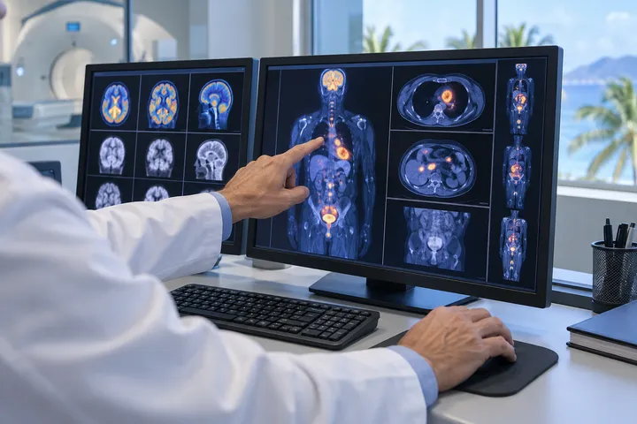
Organ and system function can be evaluated at the molecular level, often before structural changes become visible.
LIVMO — LIVE MOLECULAR IMAGES
WHAT IS LIVMO?
A functional view of the body that can reveal change earlier and support more informed care.
01
Molecular Images
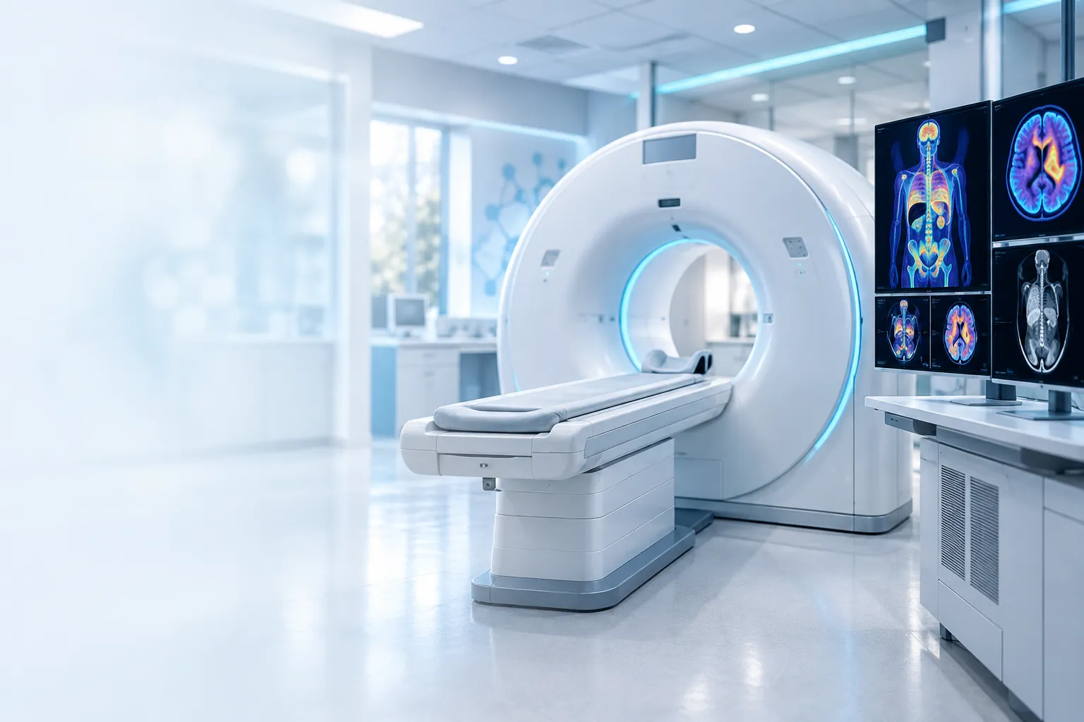
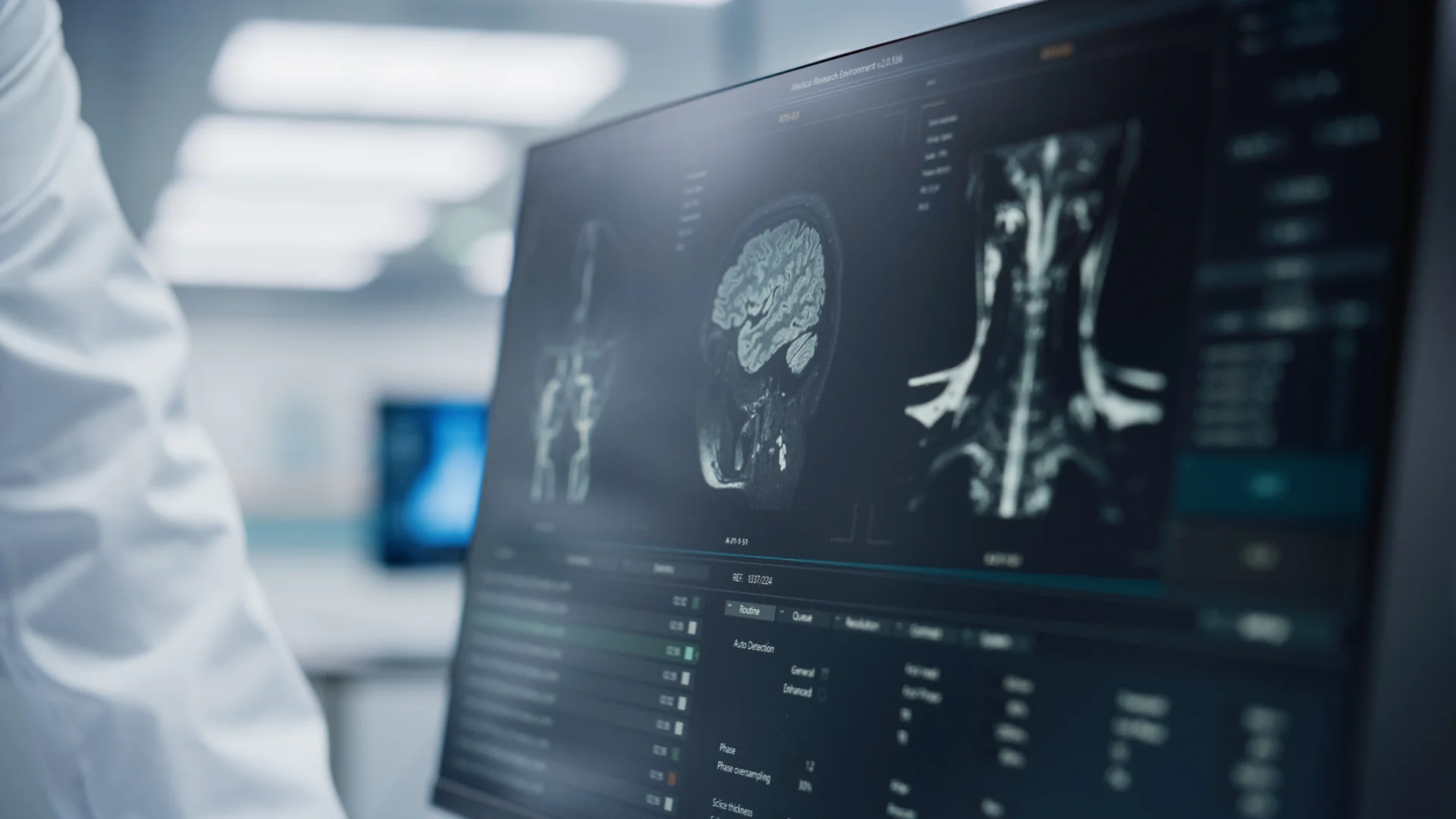
Molecular images show what is happening inside the organs, cells, and molecules of living bodies. They reveal how organs function and can support earlier diagnoses.
Beyond Anatomy
Molecular images provide detailed pictures of the activity taking place inside the organs, cells, and molecules of living bodies. Unlike techniques that primarily show structure and size, Molecular Imaging helps physicians understand how organs are functioning.
Earlier Detection
An organ such as the heart or gallbladder may have a normal size, location, and shape without functioning normally. Because functional changes usually occur before structural changes, Molecular Imaging can support earlier diagnosis when many diseases or conditions may still be treated.
Supporting Better Decisions
In coronary artery disease, Molecular Imaging can help identify which area of the heart is most affected. When an obstruction is severe, this information can help physicians evaluate whether coronary surgery may prevent permanent damage to the heart muscle.
02
Nuclear Medicine
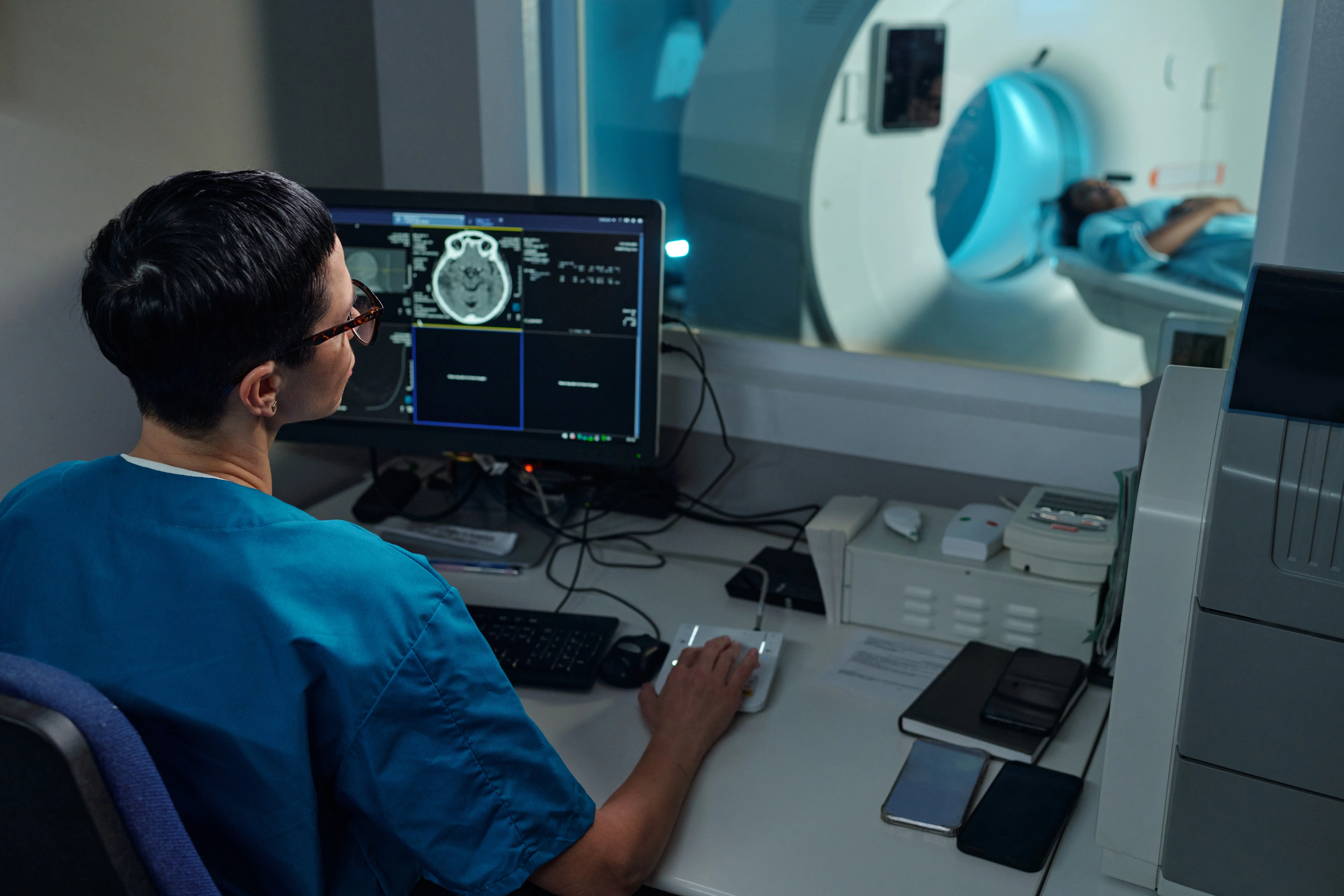
Nuclear Medicine, also known as Molecular Imaging, uses small amounts of radiopharmaceuticals to show how organs and biological processes function, often before symptoms appear.
How It Works
What has been known for decades as Nuclear Medicine is also Molecular Imaging. The amount of radiopharmaceutical administered is minimal and is directed toward the cells that need to be observed and studied. These techniques allow physicians to see how an organ or organ system works and to measure biological processes, such as heart muscle function or gallbladder contraction.
Insight Before Symptoms
We can also detect problems in organ and system function before a patient develops symptoms. This often occurs in cancer care, when very small tumors can be identified even when the patient has no symptoms, either before or after treatment.
03
Molecular Medicine
Molecular Medicine helps personalize treatment by revealing disease severity, tumor spread, response to therapies, and changes in organ function.
Personalized Treatment Insights
Molecular Medicine gives us access to bodily functions that can help personalize patient treatment. It can show disease severity, the extent of tumors, a patient's response to different therapies, and disease progression.
Measuring Treatment Response
It can also help measure how organs respond after a specific treatment, including improvement in kidney function after the removal of obstructing kidney stones, cardiac function after different types of surgery, and liver function after a patient stops consuming excessive amounts of alcohol.
INFORMED CARE
How Can It Help Me?
If we can see abnormalities in the function of our organs and systems while we are still strong and can undergo treatment with medication or surgery, the outcomes of those treatments may be better and we may be able to remain healthier for a longer time.
In addition, when information about blood changes, organ function, and symptoms is integrated, patients and a variety of specialists can collaborate to decide which treatment may be best and even integrate alternative methods, such as acupuncture, into therapeutic plans. In certain cases, when cure cannot be attempted, palliative treatments may help maintain quality of life instead of using aggressive therapy when a disease is at a very advanced stage.
OUR FOUNDER
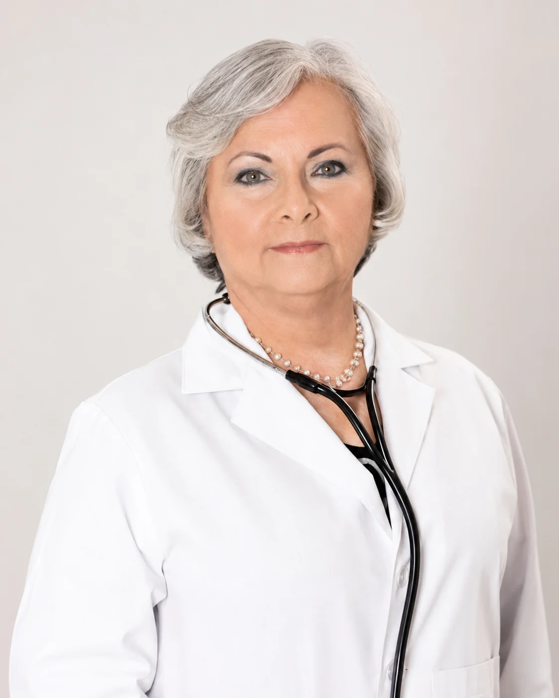
Sandra C. Gracia López, MD, DABNM, DABNC
Dr. Sandra Gracia López has more than 30 years of experience in Nuclear Medicine and an outstanding record of achievement and recognition in both the public and private sectors, including the University of Puerto Rico School of Medicine and, more recently, three Mennonite Hospital facilities in Puerto Rico.
ABOUT US
A team of experienced experts in the field.
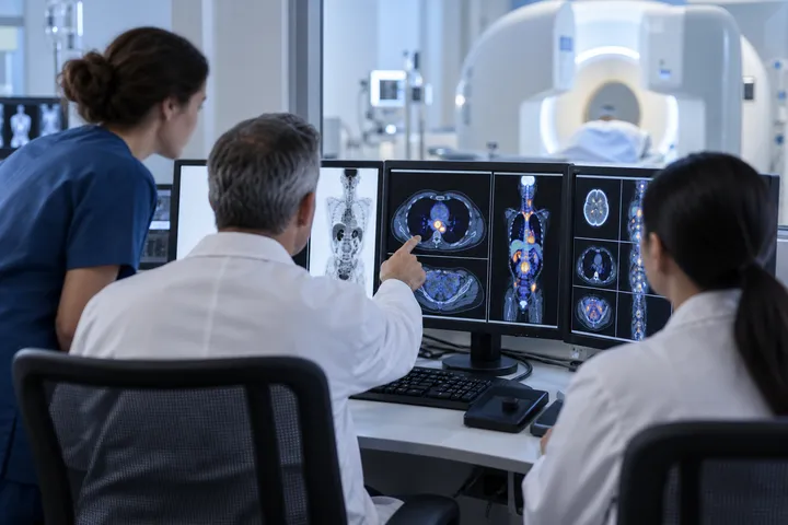
We are an integrated team of professionals certified by the American Board of Nuclear Medicine Technology, the American Board of Nuclear Medicine, and the American Board of Nuclear Cardiology, with extensive experience in the specialty. Some team members have 27 years of experience in Internal Medicine and Nuclear Medicine/Live Molecular Imaging and are board-certified in Nuclear Medicine and Nuclear Cardiology. Our team also includes cardiologists, a physicist, and administrative personnel with excellent customer service skills. We have expertise in radiation protection and extensive training and experience in developing exercises and organizing drills related to radiation incidents, accidents, and consequence management.
Our team has earned certifications from The Joint Commission and the Intersocietal Accreditation Council in Nuclear Medicine, Nuclear Cardiology, and PET/CT. Recognized by local and international patients for our excellent customer service, our goal is to help patients and referring physicians have a positive experience and make better-informed diagnostic and treatment decisions.
27+ Years of Experience
Internal Medicine and Nuclear Medicine
Board-Certified Team
Nuclear Medicine and Nuclear Cardiology
Radiation Protection
Specialized training and experience
Patient-Centered Service
Supporting patients and referring physicians
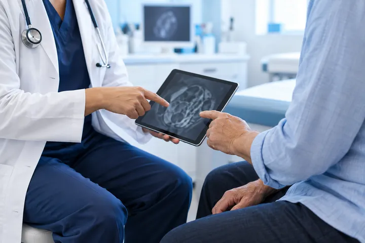
Editorial images are illustrative and do not depict LIVMO personnel or patients.
Explore our resources
Learn more about LivMo and the evolution of medical imaging.